Are you looking for an answer to the topic “How is Bifascicular block diagnosed?“? We answer all your questions at the website Chiangmaiplaces.net in category: +100 Marketing Blog Post Topics & Ideas. You will find the answer right below.
Tests that can be used to diagnose a bundle branch block or its causes include: Electrocardiogram. This records the electrical impulses in your heart through wires attached to the skin on your chest and other places on your body.Bifascicular blocks of this type are potentially significant because they make ventricular conduction dependent on the single remaining fascicle. Additional damage to this third remaining fascicle may completely block AV conduction, producing third-degree heart block (the most severe form of trifascicular block).The criteria to diagnose a LAFB, or LAHB, on ECG include the following: Left axis deviation of at least -45 degrees. The presence of a qR complex in lead I and a rS complex in lead III. Usually a rS complex in lead II and III (sometimes aVF as well)
- Conduction to the ventricles is via the single remaining fascicle.
- The ECG will show typical features of RBBB plus either left or right axis deviation.
- RBBB + LAFB is the most common of the two patterns.
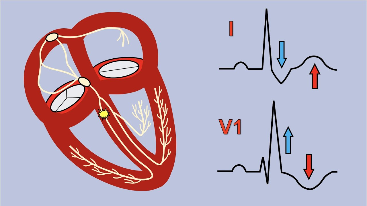
Table of Contents
How can you tell if you have a bifascicular block?
- Conduction to the ventricles is via the single remaining fascicle.
- The ECG will show typical features of RBBB plus either left or right axis deviation.
- RBBB + LAFB is the most common of the two patterns.
Is bifascicular block serious?
Bifascicular blocks of this type are potentially significant because they make ventricular conduction dependent on the single remaining fascicle. Additional damage to this third remaining fascicle may completely block AV conduction, producing third-degree heart block (the most severe form of trifascicular block).
Right Bundle Branch Block and Bifascicular Block
Images related to the topicRight Bundle Branch Block and Bifascicular Block
How is a diagnosis of a Fascicular block determined?
The criteria to diagnose a LAFB, or LAHB, on ECG include the following: Left axis deviation of at least -45 degrees. The presence of a qR complex in lead I and a rS complex in lead III. Usually a rS complex in lead II and III (sometimes aVF as well)
What causes Bifascicular heart block?
A bifascicular block can occur as a part of the ischemic heart disease or as a part of the normal degeneration of the conduction system (Lev’s disease).
What is the treatment for a bifascicular block?
Bifascicular block treatment
If you have a heart condition causing bundle branch block, treatment might involve medications to reduce high blood pressure or lessen the effects of heart failure. Additionally, depending on your symptoms and whether you have other heart problems, your doctor might recommend: A pacemaker.
How is bifascicular block treated?
In those with bifascicular block and no symptoms, little with respect to treatment is needed. In those with syncope, a pacemaker is recommended.
Does bifascicular block emergency?
Implications: o In an asymptomatic patient, bifascicular block is largely incidental and no workup is indicated. o If the patient presents with syncope and bifascicular block, this is a medical emergency and the American College of Cardiology and AHA recommend pacemaker implantation.
See some more details on the topic How is Bifascicular block diagnosed? here:
Chronic bifascicular blocks – UpToDate
Bifascicular block, a pattern seen on the surface electrocardiogram (ECG), results when normal physiologic activation in the His-Purkinje …
Bundle branch block – Diagnosis and treatment – Mayo Clinic
If you have bundle branch block with low heart-pumping function, you may need cardiac resynchronization therapy (biventricular pacing). This …
Bifascicular Block • LITFL • ECG Library Diagnosis
A new-onset bifascicular block in the context of chest pain is highly associated with proximal LAD occlusion, even in the absence of ST-segment …
Bifascicular Block: Causes, Symptoms & Treatment
What causes a bifascicular block? · Heart attack (myocardial infarction). · Heart (cardiovascular) disease. · Heart valve disease. · High potassium …
Is bifascicular block reversible?
Presence or absence of heart disease is typically based on the results of echocardiogram, coronary angiography and/or cardiac MRI. Unfortunately LBBB is not reversible.
How is heart block detected?
An electrocardiogram (ECG) is the main test used to diagnose heart block. It measures the electrical activity of your heart. An ECG can be carried out at rest or while you’re exercising. Your doctor may ask you to wear a portable ECG monitor to get a reading over time.
Can you live a normal life with left bundle branch block?
In young and healthy people, left bundle branch block is rare. This condition seems to have little effect on how long you live if you have no other underlying heart problems. You may not need any treatment at all, . especially when you have no other disease affecting your heart.
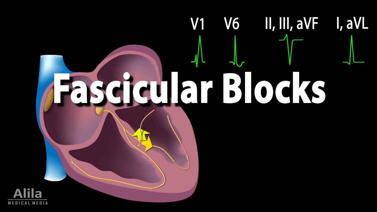
Fascicular Blocks, Animation
Images related to the topicFascicular Blocks, Animation
What does LAFB look like on EKG?
Typical ECG of LAFB, demonstrating: rS complexes in leads II, III, aVF, with small R waves and deep S waves. qR complexes in leads I, aVL, with small Q waves and tall R waves. Left Axis Deviation (LAD): Leads II, III and aVF are NEGATIVE; Leads I and aVL are POSITIVE.
Is LAFB life threatening?
Background. Right bundle branch block (RBBB) and left anterior fascicular block (LAFB) are very common findings. In the presence of second degree atrio-ventricular (AV) block, this condition can be life threatening and mandates emergent treatment.
Is RBBB life threatening?
In people with known or suspected heart disease, right bundle branch block is associated with a greater risk of death, especially after a heart attack. Some people with right bundle branch block may ultimately need a permanent pacemaker, but this is rare.
What is Bifascicular?
● Bifascicular block – The term bifascicular block most commonly refers to conduction disturbances below the atrioventricular (AV) node in which the right bundle branch and one of the two fascicles (anterior or posterior) of the left bundle branch are involved.
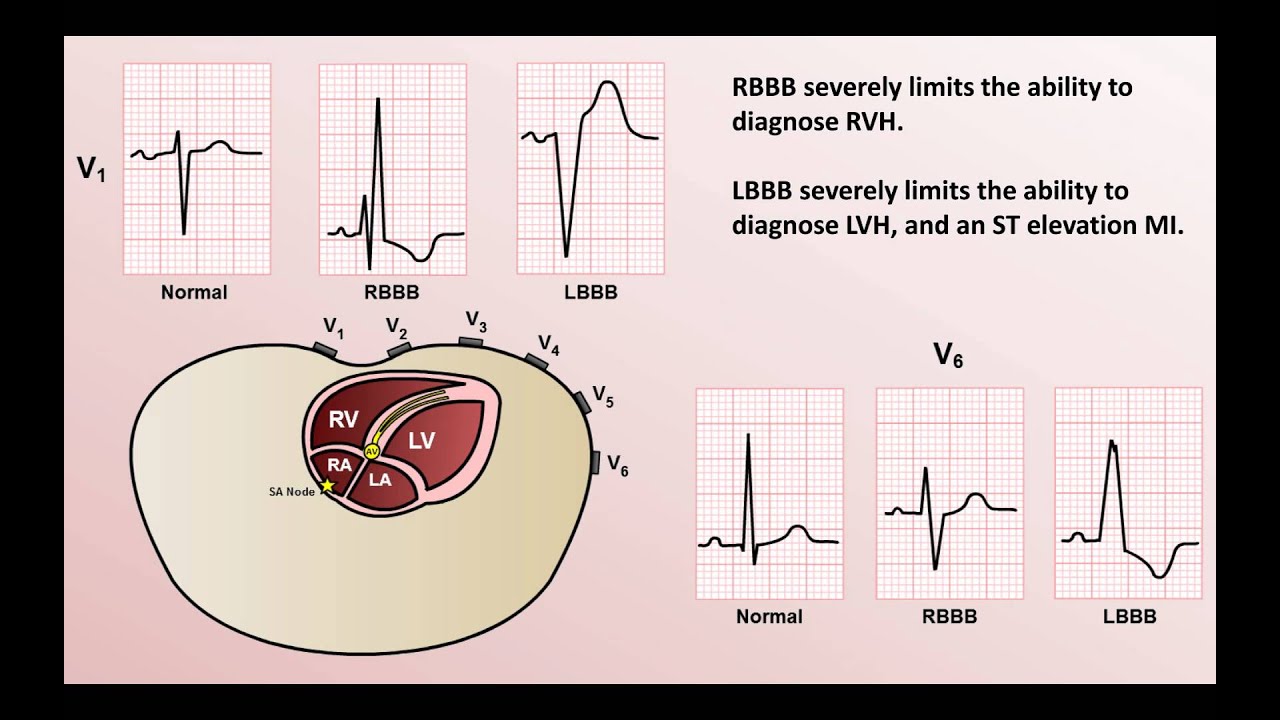
How can you tell if you have a bundle branch block on ECG?
- QRS duration greater than 120 milliseconds.
- rsR’ “bunny ear” pattern in the anterior precordial leads (leads V1-V3)
- Slurred S waves in leads I, aVL and frequently V5 and V6.
What medication is given for heart block?
Medications that may be used in the management of third-degree AV block (complete heart block) include sympathomimetic or vagolytic agents, catecholamines, and antidotes.
Can bifascicular block cause syncope?
Introduction. Bifascicular block (BFB) is a conduction disturbance with reported prevalence of 1% to 1.5%, with up to 25% of adult patients presenting with syncope.
Can I exercise with heart block?
Get regular exercise. Try for 2½ hours a week. If you do not have other heart problems, you likely do not have limits on the type or level of activity that you can do. You may want to walk, swim, bike, or do other activities.
Can first degree heart block get worse?
In rare instances, a first-degree heart block may develop into a more serious type of heart block that results in slower heartbeats. This may cause symptoms.
Intro to EKG Interpretation – Bundle Branch Blocks
Images related to the topicIntro to EKG Interpretation – Bundle Branch Blocks
How long can you live with right bundle branch block?
If you don’t have heart disease, having right bundle branch block doesn’t change your life expectancy or add to your risk level. But having right bundle branch block can put you at a higher risk of death if you also have heart failure or a heart attack.
What is the ICD 10 code for bifascicular block?
ICD-10 | Bifascicular block (I45. 2)
Related searches to How is Bifascicular block diagnosed?
- is bifascicular block reversible
- how serious is bifascicular block
- how is bifascicular block diagnosed
- is bifascicular block dangerous
- bifascicular block life expectancy
- bifascicular block treatment
- bifascicular block criteria
- bifascicular block and beta blockers
- bifascicular block pacemaker
- how do you treat a bifascicular block
- living with bifascicular block
- bifascicular block symptoms
- what is the treatment for bifascicular block
Information related to the topic How is Bifascicular block diagnosed?
Here are the search results of the thread How is Bifascicular block diagnosed? from Bing. You can read more if you want.
You have just come across an article on the topic How is Bifascicular block diagnosed?. If you found this article useful, please share it. Thank you very much.