Are you looking for an answer to the topic “How is hydatidiform mole diagnosed?“? We answer all your questions at the website Chiangmaiplaces.net in category: +100 Marketing Blog Post Topics & Ideas. You will find the answer right below.
Ultrasonography is done to be sure that the growth is a hydatidiform mole and not a fetus or amniotic sac (which contains the fetus and fluid around it). (D and C) or obtained when tissue is passed and is then examined under a microscope (biopsy) to confirm the diagnosis.The most common symptom (in one study as high as 84% of patients) of a complete mole is vaginal bleeding in the first trimester, which is normally due to the molar tissue separating from the decidua, resulting in bleeding.There are often no symptoms of a molar pregnancy. It may only be diagnosed during a routine ultrasound scan at 8-14 weeks or during tests are done after a miscarriage.
- Dark brown to bright red vaginal bleeding during the first trimester.
- Severe nausea and vomiting.
- Sometimes vaginal passage of grapelike cysts.
- Pelvic pressure or pain.

Table of Contents
What is the most common manifestation of a patient diagnosed with hydatidiform mole?
The most common symptom (in one study as high as 84% of patients) of a complete mole is vaginal bleeding in the first trimester, which is normally due to the molar tissue separating from the decidua, resulting in bleeding.
What are the signs and symptoms of hydatidiform mole?
- Dark brown to bright red vaginal bleeding during the first trimester.
- Severe nausea and vomiting.
- Sometimes vaginal passage of grapelike cysts.
- Pelvic pressure or pain.
Hydatidiform Mole
Images related to the topicHydatidiform Mole
When can a molar pregnancy be detected?
There are often no symptoms of a molar pregnancy. It may only be diagnosed during a routine ultrasound scan at 8-14 weeks or during tests are done after a miscarriage.
How is partial molar pregnancy diagnosed?
How is a partial molar pregnancy diagnosed? The doctor diagnoses a molar pregnancy by performing an ultrasound, which will reveal the presence of cysts in the uterus. The doctor will also perform another test that measures the levels of beta human chorionic gonadotropin (hCG).
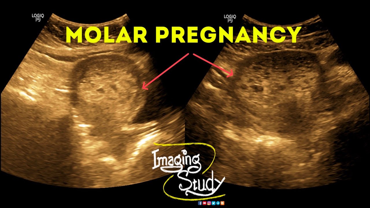
What does a molar pregnancy look like on ultrasound?
The presence of the molar tissue is then detected. Ultrasound scanning shows a honeycomb pattern produced by the numerous vesicles. As they enlarge the image is described to look like a snowstorm, which is due to swollen cysts with bleeding into the uterus. The ovaries are often seen to contain large cysts.
How do they remove a molar pregnancy?
Treatment usually consists of one or more of the following steps: Dilation and curettage (D&C). To treat a molar pregnancy, your doctor will remove the molar tissue from your uterus with a procedure called dilation and curettage ( D&C ). A D&C is usually done as an outpatient procedure in a hospital.
Will a molar pregnancy test positive?
Women with a molar pregnancy will have a positive pregnancy test and the same early symptoms of a normal pregnancy. In the absence of medical intervention or diagnosis, the pregnancy might seem normal for the first three to four months.
See some more details on the topic How is hydatidiform mole diagnosed? here:
Molar pregnancy – Diagnosis and treatment – Mayo Clinic
If your doctor suspects a molar pregnancy, he or she will order blood tests, including one to measure the level of human chorionic …
Hydatidiform mole: Epidemiology, clinical features … – UpToDate
Routine pre-evacuation ultrasound diagnosis of hydatidiform mole: experience of more than 1000 cases from a regional referral center.
Hydatidiform mole | The Royal Women’s Hospital
A hydatidiform mole is sometimes detected when you have an early pregnancy ultrasound. It may also be diagnosed after a miscarriage, when the tissue that is …
Hydatidiform Mole – Medscape Reference
Diagnosis of hydatidiform mole · Quantitative beta-human chorionic gonadotropin (hCG) levels · Complete blood cell count with platelets · Clotting …
What is the medical management for H mole?
Treatment involves surgical removal of the molar pregnancy followed by surveillance of serial human chorionic gonadotropin (hCG) levels to confirm resolution of disease or to identify development of gestational trophoblastic neoplasia (GTN), which includes invasive mole, choriocarcinoma, placental site trophoblastic …
Is there a heartbeat in molar pregnancy?
Diagnosis. Most molar pregnancies are diagnosed in the first trimester. This condition may be discovered when a heartbeat does not become detectable by 12 weeks, but this can also be true of missed miscarriages.
Molar Pregnancy
Images related to the topicMolar Pregnancy

How high are hCG levels in a molar pregnancy?
The measurement of high hCG levels in excess of 100,000 mIU/mL suggests the diagnosis of a complete molar pregnancy, particularly when associated with vaginal bleeding, uterine enlargement and abnormal ultrasound findings.
How quickly do hCG levels drop after molar pregnancy?
If the levels of a hormone called hCG go back to normal soon after removal of the molar pregnancy then your doctor won’t need to give it a stage. In most women, the hCG level virtually disappears within 4 to 6 weeks of removing the molar pregnancy.
How is pregnancy diagnosed?
Currently, most women are diagnosed with pregnancy after a missed menstrual cycle and a positive urine or serum hCG. The pregnancy is diagnosed as viable with serial exams and normal pregnancy development, a normal dating ultrasound, or positive fetal heart tones by Doppler.
What should be the hCG level at 5 weeks?
At 5 weeks pregnant, your hCG levels can range from about 217 to 8,245 mIU/mL.
What are hCG levels in pregnancy?
An hCG level of less than 5 mIU/mL is considered negative for pregnancy, and anything above 25 mIU/mL is considered positive for pregnancy. An hCG level between 6 and 24 mIU/mL is considered a grey area, and you’ll likely need to be retested to see if your levels rise to confirm a pregnancy.
Can you naturally miscarry a molar pregnancy?
A molar pregnancy will not be able to survive. It may end on its own, with a miscarriage. If this does not happen, it’s usually treated with a procedure to remove the pregnancy.
Does a molar pregnancy have a yolk sac?
Molar pregnancy ultrasound
In a healthy pregnancy, your doctor would point out the gestational sac, the yolk sac, and the fetal pole at 9 weeks. In a complete molar pregnancy, these structures are absent and there’s only abnormal placental tissue that fills the uterine cavity.
What percent of molar pregnancies are cancerous?
Most molar pregnancies are mostly benign (not cancerous). They are rare but they are the most common type of gestational trophoblastic tumour. In the UK, about 1 in 590 pregnancies is a molar pregnancy. In Asian women molar pregnancies are about twice as common as in Caucasian women.
Molar Pregnancy || Hydatidiform Mole || Gestational Trophoblastic Disease || Ultrasound || Case 38
Images related to the topicMolar Pregnancy || Hydatidiform Mole || Gestational Trophoblastic Disease || Ultrasound || Case 38
Can IVF prevent molar pregnancy?
For IVF patients, a PGD-tested embryo can virtually eliminate the risk of a molar pregnancy.
Are all molar pregnancies cancerous?
A molar pregnancy contains many cysts (sacs of fluid). It is usually benign (not cancer) but it may spread to nearby tissues (invasive mole). It may also become a malignant tumor called choriocarcinoma. Molar pregnancy is the most common type of gestational trophoblastic tumor.
Related searches to How is hydatidiform mole diagnosed?
- complications of hydatidiform mole
- how is hydatidiform mole diagnosis
- hydatidiform mole pictures
- partial hydatidiform mole
- hydatidiform mole treatment
- how does hydatidiform mole occur
- bleeding after molar pregnancy removal
- molar pregnancy hcg levels chart
- complete hydatidiform mole
- hydatidiform mole symptoms
- is hydatidiform mole hereditary
- hydatidiform mole differential diagnosis
Information related to the topic How is hydatidiform mole diagnosed?
Here are the search results of the thread How is hydatidiform mole diagnosed? from Bing. You can read more if you want.
You have just come across an article on the topic How is hydatidiform mole diagnosed?. If you found this article useful, please share it. Thank you very much.